What Is PAH-CTD?

PAH Is a Serious Complication of CTD1

Pulmonary arterial hypertension (PAH, WHO Group I) is a progressive syndrome resulting from restricted blood flow through the pulmonary arterial circulation.2

PAH LEADS TO2:

PAH LEADS TO2:

  • Reduced cardiac output
  • Reduced cardiac output
  • Right heart failure
  • Right heart failure
  • Death
  • 30% mortality rate at 3 years from diagnosis observed in the REVEAL Registry
  • Death
  • 30% mortality rate at 3 years from diagnosis observed in the REVEAL Registry

PAH SUBGROUPS INCLUDE3:

PAH SUBGROUPS INCLUDE3:

  • Idiopathic PAH
  • Idiopathic PAH
  • Heritable PAH
  • Heritable PAH
  • Drug- or toxin-induced PAH
  • Drug- or toxin-induced PAH
  • PAH associated with other diseases or conditions, including connective tissue disease (CTD)

Prevalence of PAH-SSc

PAH associated with connective tissue disease (PAH-CTD) is the second most prevalent type of PAH (after idiopathic PAH) in Western countries.3

PAH-CTD is more prevalent in women than men (4:1 ratio)3

PAH may be a serious complication of CTD such as3:

  • Systemic sclerosis (SSc)
  • Systemic sclerosis (SSc)
  • Mixed CTD
  • Mixed CTD
  • Systemic lupus erythematosus (SLE)
  • Systemic lupus erythematosus (SLE)
PAH-CTD associations1
PAH-CTD associations chart PAH-CTD associations chart

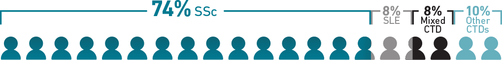
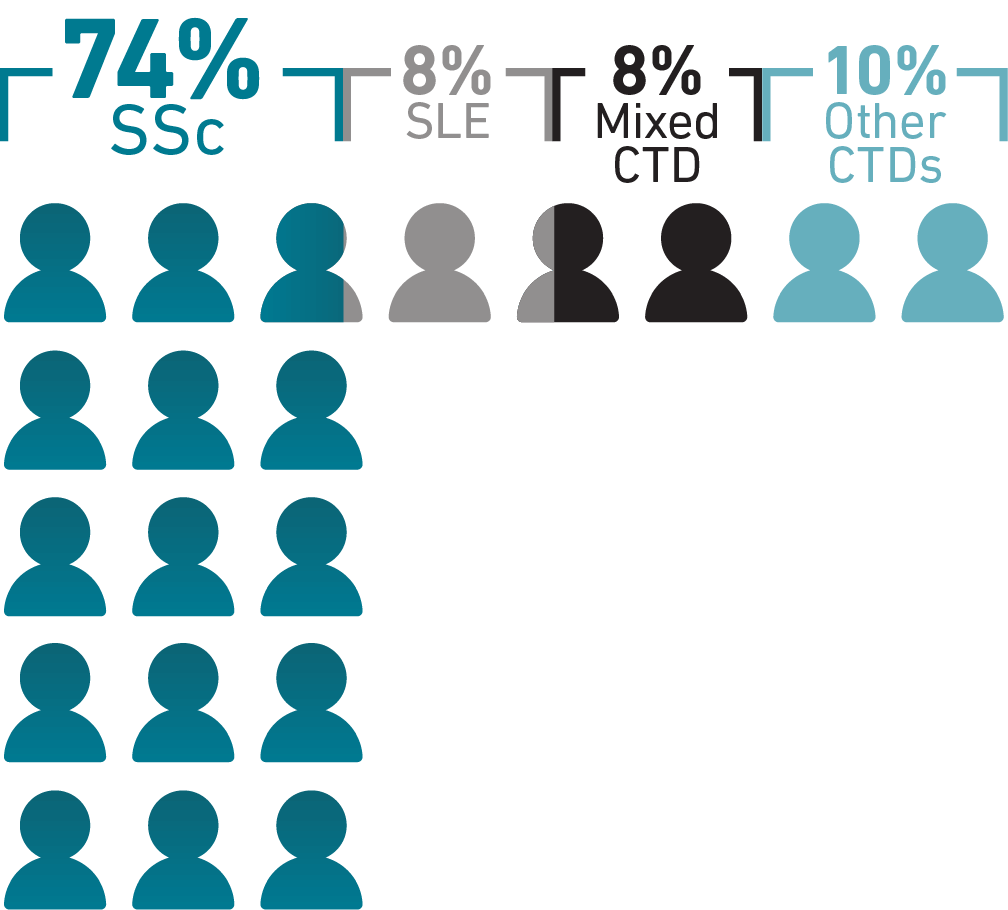
While SLE and rheumatoid arthritis are among the most common CTDs, PAH occurs most 
frequently in SSc, with 74% of PAH-CTD cases being diagnosed in patients with SSc.1

~100,000 people living with SSc in the United States4
In DETECT, an international study of adults with SSc at increased risk of PAH (N=466), 19% were confirmed to have PAH-SSc5*
In the DETECT study,44% of patients with PAH-SSc with follow-up data (25/57) experienced disease progression within a short period of time (median observation time, 12.6 months)6*
You can help make the connection between CTD and the risk of PAH earlier using noninvasive tests—see how the DETECT algorithm can help identify patients with SSc in need of right heart catheterization.5 Learn More >

PAH-CTD in African American Patients

PAH-CTD in African American Patients

Image
Image

PAH-CTD may disproportionately occur in African Americans
compared with other races.7 That disparity may persist in PAH-SSc:

African American patients were 2.5 times more likely to have PAH-CTD than non-Hispanic White and Hispanic patients in the National Biological Sample and Data Repository for PAH (PAH Biobank)7†

In a 2003 single-center US study, 48% of African American patients in a scleroderma center (n=130) had PAH, compared with 39% overall (n=709)8

Another study reported that African American patients (n=29) had greater disease severity at PAH-SSc diagnosis, including worse FC, right ventricular function, NT-proBNP, and 6-minute walk distance than European American patients (n=131)9

In 2 studies using national registries of death certificate data, African American women had the highest mortality rates of patients with PAH

Pathogenesis of PAH

SSc is a chronic, inflammatory, autoimmune disease characterized by endothelial dysfunction and microvascular disease.10-12 This baseline inflammation and endothelial dysfunction contributes to the development of associated PAH, followed by vasoconstriction and vascular remodeling and eventual right heart failure.1,10

CHANGES IN PULMONARY ARTERY
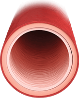
Image
HEALTHY ARTERY
Healthy endothelium regulates13:
  • Vasodilation/vasoconstriction balance
  • Inhibited smooth muscle cell proliferation
  • Low-resistance vasculature
Image
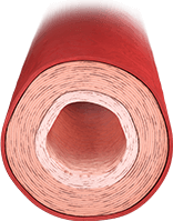
ARTERY WITH EFFECTS FROM PAH
  • Earlier-stage PAH is characterized by smooth muscle hypertrophy and vasoconstriction14
Endothelial dysfunction results in15:
  • Overproduction of vasoconstrictors such as endothelin-1
  • Underproduction of vasodilators such as nitric oxide and prostacyclin
Image
ARTERY WITH WORSENING SIGNS OF PAH
Proliferative features predominate
in later stages14:
  • Increased vasoconstriction
  • Obstruction from plexiform lesions and in situ thrombosis

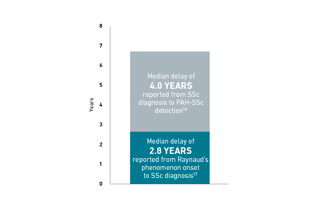
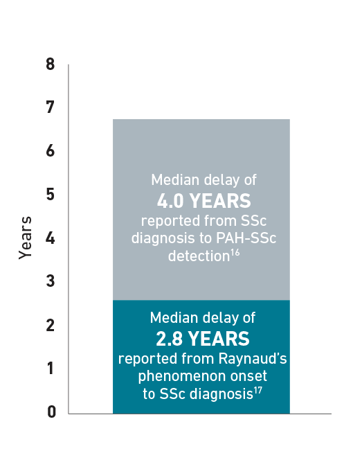
Delayed Diagnosis of PAH-SSc

Patients with SSc can develop PAH at any time, but experience a median delay in diagnosis of 4 years.16 The following chart is an example for illustrative purposes only to portray the delay in diagnosis of PAH-SSc that a patient may experience. In some patients with SSc, median time to diagnosis of SSc after Raynaud’s phenomenon onset was 2.8 years.17 Additionally, median time to diagnosis of PAH in patients with SSc was 4 years.16

Delay in PAH-SSc diagnosis16,17

4-year chart showing PAH-CTD diagnosis to detection
Image
Learn about annual screenings for PAH in patients with SSc.3 Find Out What the Guidelines Say >

Symptoms and Risk Factors

Awareness of symptoms and predictive risk factors can help identify PAH in patients. Misdiagnosis and delayed diagnosis may be attributed to the nonspecific nature of the presenting symptoms.18,19

Symptoms of PAH-SSc10

Woman staring into the distance with arms folded Image

Dyspnea

Woman staring into the distance with arms folded Image

Fatigue

Woman staring into the distance with arms folded Image

Orthostasis

Woman staring into the distance with arms folded Image

Chest pain/angina

Woman staring into the distance with arms folded Image

Lower extremity edema

Woman staring into the distance with arms folded Image

Syncope

Regular screening followed by diagnostic right heart catheterization is required to consistently detect PAH-SSc.20

Risk factors for PAH-SSc21,22

Time-Related Risk Factors

  • Older age at scleroderma onset
  • Long-standing disease

Coexisting Conditions

  • Limited cutaneous scleroderma
  • Cutaneous telangiectasias
  • Severe digital ischemia
  • Severe Raynaud’s phenomenon

Diagnostic Results

  • Nucleolar pattern of antinuclear antibodies
  • Anticentromere antibodies
  • Declining or isolated low Dlco
  • Low FVC %/ Dlco %
*DETECT was a global cross-sectional study involving 62 experienced centers. DETECT was funded and supported by Actelion Pharmaceuticals Ltd. 488 patients with SSc enrolled between 2008 and 2011, and nearly 1 in 5 (19%, 87/466) were found to have PAH. Disease progression was defined as FC worsening, combination therapy for PAH, PAH-related hospitalization, or death.5,6
This 2017 study of racial and ethnic differences in PAH included 1837 White, African American, and Hispanic patients enrolled in PAH Biobank.7
Racial classification was based on self-report. Hispanic patients (2% of 131) were considered together with White patients within the group of European Americans for simplicity.9
§In an analysis of 5960 patients, the mortality rate was 3x higher in African American women (n=1306) compared with Caucasian women (n=4654). An increased risk of death (HR 3.20 [95% CI: 1.3-7.6]) for patients of Asian or African-American descent was also reported in a separate analysis of 84 patients with PAH. In this population of 69 non-Hispanic White and Hispanic, 9 Asian, and 6 African American patients, 81% were women.23,24
CI=confidence interval; CTD=connective tissue disease; Dlco=diffusing capacity of the lungs for carbon monoxide; FC=Functional Class; FVC=forced vital capacity; HR=hazard ratio; NT-proBNP=N-terminal pro-brain natriuretic peptide; PAH=pulmonary arterial hypertension; PAH-CTD=pulmonary arterial hypertension associated with CTD; SLE=systemic lupus erythematosus; SSc=systemic sclerosis; WHO=World Health Organization.
References: 1. Zanatta E, Polito P, Famoso G, et al. Pulmonary arterial hypertension in connective tissue disorders: pathophysiology and treatment. Exp Biol Med. 2019;244(2):120-131. 2. Lai Y-C, Potoka KC, Champion HC, et al. Pulmonary arterial hypertension: the clinical syndrome. Circ Res. 2014;115(1):115-130. 3. Humbert M, Kovacs G, Hoeper MM, et al; ESC/ERS Scientific Document Group. 2022 ESC/ERS Guidelines for the diagnosis and treatment of pulmonary hypertension: the Task Force for the Diagnosis and Treatment of Pulmonary Hypertension of the European Society of Cardiology (ESC) and the European Respiratory Society (ERS). Eur Heart J. 2022;43(38):3618-3731. 4. Varga J. Systemic sclerosis (scleroderma) and related disorders. In: Fauci AS, Kasper DL, Longo DL, et al, eds. Harrison’s Principles of Internal Medicine. 17th ed. McGraw-Hill; 2008:2096-2106. 5. Coghlan JG, Denton CP, Grünig E, et al; DETECT study group. Evidence-based detection of pulmonary arterial hypertension in systemic sclerosis: the DETECT study. Ann Rheum Dis. 2014;73(7):1340-1349. 6. Mihai C, Antic M, Dobrota R, et al. Factors associated with disease progression in early-diagnosed pulmonary arterial hypertension associated with systemic sclerosis: longitudinal data from the DETECT cohort. Ann Rheum Dis. 2018;77(1):128-132. 7. Al-Naamani N, Paulus JK, Roberts KE, et al. Racial and ethnic differences in pulmonary arterial hypertension. Pulm Circ. 2017;7(4):793-796. 8. Schachna L, Wigley FM, Chang B, et al. Age and risk of pulmonary arterial hypertension in scleroderma. CHEST. 2003;124(6):2098-2104. 9. Blanco I, Mathai S, Shafiq M, et al. Severity of systemic sclerosis-associated pulmonary arterial hypertension in African Americans. Medicine (Baltimore). 2014;93(5):177-185. 10. Chaisson NF, Hassoun PM. Systemic sclerosis-associated pulmonary arterial hypertension. CHEST. 2013;144(4):1346-1356. 11. Kelemen BW, Mathai SC, Tedford RJ, et al. Right ventricular remodeling in idiopathic and scleroderma-associated pulmonary arterial hypertension: two distinct phenotypes. Pulm Circ. 2015;5(2):327-334. 12. Mathai SC, Hassoun PM. Pulmonary arterial hypertension in connective tissue diseases. Heart Fail Clin. 2012;8(3):413-425. 13. Huertas A, Guignabert C, Barberá JA, et al. Pulmonary vascular endothelium: orchestra conductor in respiratory diseases: highlights from basic research to therapy. Eur Respir J. 2018;51(4);1700745. 14. Gaine S. Pulmonary hypertension. JAMA. 2000;284(24):3160-3168. 15. Minai OA, Budev MM. Diagnostic strategies for suspected pulmonary arterial hypertension: a primer for the internist. Cleve Clin J Med. 2007;74:737-747. 16. Hachulla E, Launay D, Mouthon L, et al; French PAH-SSc Network. Is pulmonary arterial hypertension really a late complication of systemic sclerosis? CHEST. 2009;136(5):1211-1219. 17. Delisle VC, Hudson M, Baron M, et al; The Canadian Scleroderma Research Group. Sex and time to diagnosis in systemic sclerosis: an updated analysis of 1,129 patients from the Canadian scleroderma research group registry. Clin Exp Rheumatol. 2014;32(6 suppl 86):S10-S14. 18. Mandras SA, Ventura HO, Corris PA. Breaking down the barriers: why the delay in referral for pulmonary arterial hypertension? Ochsner J. 2016;16(3):257-262. 19. Vachiéry J-L, Gaine S. Challenges in the diagnosis and treatment of pulmonary arterial hypertension. Eur Respir Rev. 2012;21(126):313-320. 20. Denton CP, Hachulla E. Risk factors associated with pulmonary arterial hypertension in patients with systemic sclerosis and implications for screening. Eur Respir Rev. 2011;20(122):270-276. 21. Yaqub A, Chung L. Epidemiology and risk factors for pulmonary hypertension in systemic sclerosis. Curr Rheumatol Rep. 2013;15(1):302. 22. Fischer A, Bull TM, Steen VD. Practical approach to screening for scleroderma-associated pulmonary arterial hypertension. Arthritis Care Res (Hoboken). 2012;64(3):303-310. 23. Davis KK, Lilienfeld DE, Doyle RL. Increased mortality in African Americans with idiopathic pulmonary arterial hypertension. J Natl Med Assoc. 2008;100(1):69-72. 24. Kawut SM, Horn EM, Berekashvili KK, et al. New predictors of outcome in idiopathic pulmonary arterial hypertension. Am J Cardiol. 2005;95(2):199-203.